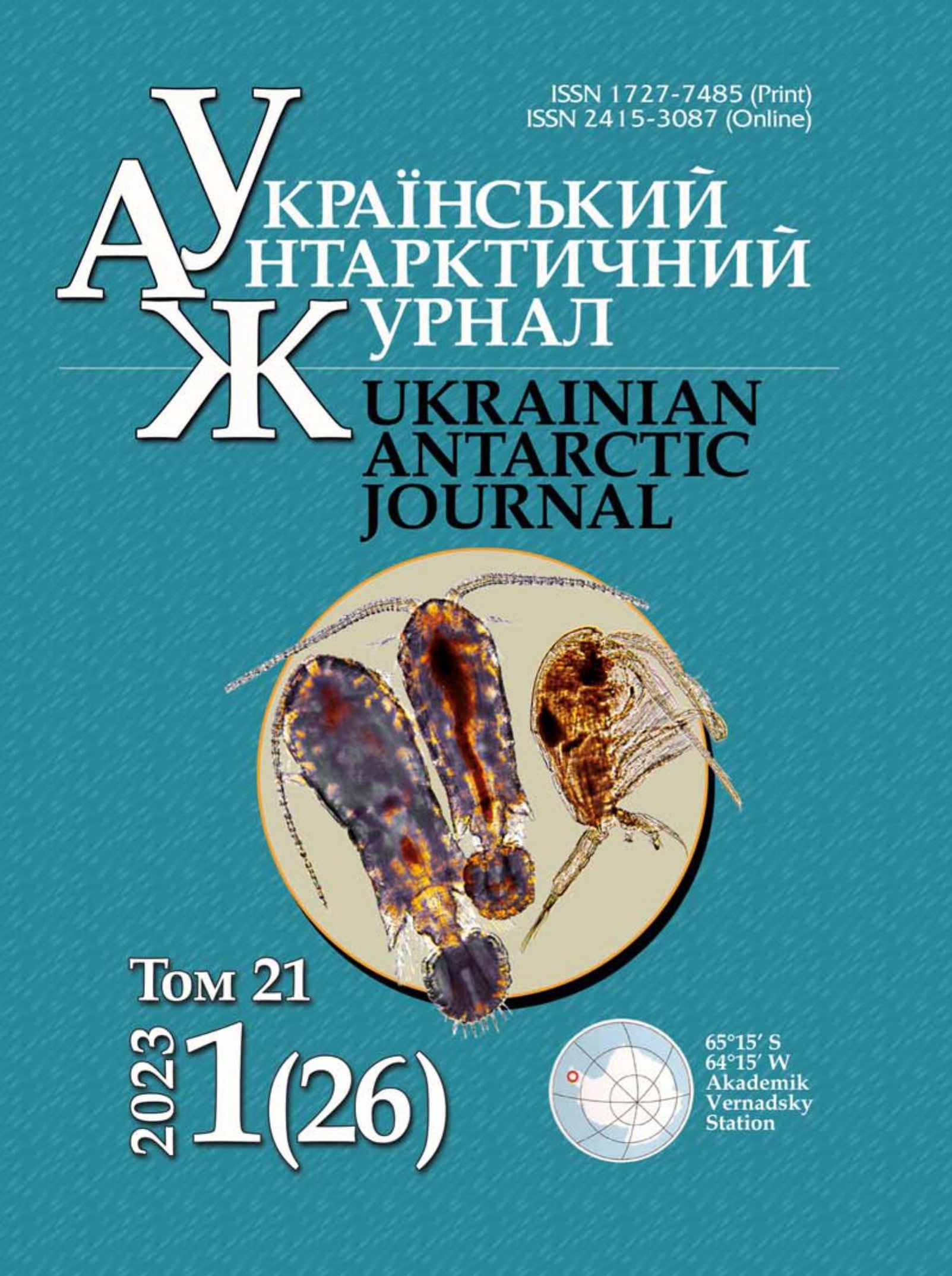
Adaptations of the antarctic bacterium Paenibacillus tundrae IMV B-7915 to copper (II) chloride exposure
- antioxidant defense,
- extracellular polymeric substances,
- free radicals,
- heavy metals,
- lipid peroxidation
- protein damage ...More
Copyright (c) 2023 Ukrainian Antarctic Journal

This work is licensed under a Creative Commons Attribution-NonCommercial-NoDerivatives 4.0 International License.
Abstract
Heavy metals are common in Antarctic habitats. However, the adaptations of Antarctic microorganisms to heavy metals are poorly understood. One of the mechanisms of toxicity of transition metals is the formation of free radicals which damage the cell macromolecules. In 2020, we isolated the bacteria Paenibacillus tundrae IMV B-7915 from a sample containing moss, soil, and underground parts of Deschampsia antarctica (Berthelot Islands, Maritime Antarctic). The aim of the study was to investigate the influence of copper (II) chloride on the specific growth rate, the content of products of free radical damage to lipids and proteins, the activity of antioxidant defense system enzymes, and the synthesis of extracellular polymeric substances in P. tundrae IMV B-7915. The bacteria were incubated for an hour in Tris-HCl buffer with 2–8 mM copper (II) chloride, then washed and inoculated into the tryptic soy broth. The bacteria were cultured for 72 hours. The content of copper in the cells was determined by atomic absorption spectrometry. The content of indicators of lipid peroxidation (diene conjugates, lipid hydroperoxides, thiobarbituric acid-reactive substances), oxidative modification of proteins (carbonyl groups in proteins), the activity of the antioxidant defense system enzymes (catalase, superoxide dismutase, glutathione peroxidase, glutathione S-transferase, glutathione reductase), total thiols, exopolymeric compounds (exopolysaccharides and proteins) were determined photometrically. Within an hour, cells accumulate 1.5–3.4 mg Cu/g of biomass, leading to a decrease in biomass accumulation and specific growth rate within 24 hours. In cells, copper ions induce free radical reactions of damage to cell macromolecules, reflected in the increase in the content of primary lipid peroxidation products and carbonyl groups in proteins. Cell division is inhibited. In response, P. tundrae IMV B-7915 cells activate efflux systems, as evidenced by a significant decrease in copper content during prolonged cultivation, and enzymes of antioxidant defense and synthesis of exopolysaccharides. The complex of the studied adaptation reactions ensures the detoxification of copper accumulated in cells, reflected in the restoration of the specific growth rate.
References
- Abdu, N., Abdullahi, A. A., & Abdulkadir, A. (2017). Heavy metals and soil microbes. Environmental Chemistry Letters, 15, 65–84. https://doi.org/10.1007/s10311-016-0587-x
- Allocati, N., Federici, L., Masulli, M., & Di Ilio, C. (2009). Glutathione transferases in bacteria. The FEBS Journal, 276(1), 58–75. https://doi.org/10.1111/j.1742-4658.2008.06743.x
- Andrei, A., Öztürk, Y., Khalfaoui-Hassani, B., Rauch, J., Marckmann, D., Trasnea, P.-I., Daldal, F., & Koch, H.-G. (2020). Cu homeostasis in bacteria: the ins and outs. Membranes, 10(9), 242. https://doi.org/10.3390/membranes10090242
- Artemenko, G. V., Ganotzkiy, V. I., Kanunikova, L. I., Grechanovskaya, E. E., & Taraschan, A. A. (2019). Occurrence of wolfram, copper, cobalt and gold mineralization in the area of the Argentine Islands (West Antarctica). Ukrainian Antarctic Journal, 2(19), 3–12. https://doi.org/10.33275/1727-7485.2(19).2019.147
- Ayala, A., Muñoz, M. F., & Argüelles, S. (2014). Lipid peroxidation: production, metabolism, and signaling mechanisms of malondialdehyde and 4-hydroxy-2-nonenal. Oxidative Medicine and Cellular Longevity, 2014, 360438. https://doi.org/10.1155/2014/360438
- Baltazar, M. D. P. G., Gracioso, L. H., Avanzi, I. R., Karol ski, B., Tenório, J. A. S., do Nascimento, C. A. O., & Perpetuo, E. A. (2019). Copper biosorption by Rhodococcus erythropolis isolated from the Sossego Mine–PA–Brazil. Journal of Materials Research and Technology, 8(1), 475–483. https://doi.org/10.1016/j.jmrt.2018.04.006
- Bedernichek, T., Loya, V., & Parnikoza, I. (2020). Content of biogenic and toxic elements in the leaves of Deschampsia antarctica É. Desv. (Poaceae): a preliminary study. Plant Introduction, 1(85/86), 124–129. https://doi.org/10.46341/PI2020017
- Bosmans, L., De Bruijn, I., Gerards, S., Moerkens, R., Van Looveren, L., Wittemans, L., Van Calenberge, B., Paeleman, A., Van Kerckhove, S., De Mot, R., Rozenski, J., Rediers, H., Raaijmakers, J. M., & Lievens, B. (2017). Potential for biocontrol of hairy root disease by a Paenibacillus clade. Frontiers in Microbiology, 8, 447. https://doi.org/10.3389/fmicb.2017.00447
- Bradford, M. M. (1976). A rapid and sensitive method for the quantitation of microgram quantities of protein utilizing the principle of protein-dye binding. Analytical Biochemistry, 72(1–2), 248–254. https://doi.org/10.1016/0003-2697(76)90527-3
- Dsouza, M., Taylor, M. W., Turner, S. J., & Aislabie, J. (2014). Genome-based comparative analyses of Antarctic and temperate species of Paenibacillus. PloS One, 9(10), e108009. https://doi.org/10.1371/journal.pone.0108009
- Ezraty, B., Gennaris, A., Barras, F., & Collet, J.-F. (2017). Oxidative stress, protein damage and repair in bacteria. Nature Reviews Microbiology, 15(7), 385–396. https://doi.org/10.1038/nrmicro.2017.26
- Frølund, B., Palmgren, R., Keiding, K., & Nielsen, P. H. (1996). Extraction of extracellular polymers from activated sludge using a cation exchange resin. Water Research, 30(8), 1749–1758. https://doi.org/10.1016/0043-1354(95)00323-1
- Fu, R.-Y., Chen, J., & Li, Y. (2007). The function of the glutathione/glutathione peroxidase system in the oxidative stress resistance systems of microbial cells. Chinese Journal of Biotechnology, 23(5), 770–775. https://doi.org/10.1016/S1872-2075(07)60048-X
- Ghaed, S., Shirazi, E. K., & Marandi, R. (2013). Biosorption of copper ions by Bacillus and Aspergillus species. Adsorption Science & Technology, 31(10), 869–890. https://doi.org/10.1260/0263-6174.31.10.869
- Gout, I. (2019). Coenzyme A: a protective thiol in bacterial antioxidant defence. Biochemical Society Transactions, 47(1), 469–476. https://doi.org/10.1042/bst20180415
- Havryliuk, O., Hovorukha, V., Patrauchan, M., Youssef, N. H., & Tashyrev, O. (2020). Draft whole genome sequence for four highly copper resistant soil isolates Pseudomonas lactis strain UKR1, Pseudomonas panacis strain UKR2, and Pseudomonas veronii strains UKR3 and UKR4. Current Research in Microbial Sciences, 1, 44–52. https://doi.org/10.1016/j.crmicr.2020.06.002
- Hawkins, C. L., & Davies, M. J. (2019). Detection, identification, and quantification of oxidative protein modifications. Journal of Biological Chemistry, 294(51), 19683–19708. https://doi.org/10.1074/jbc.REV119.006217
- Hawkins, C. L., Morgan, P. E., & Davies, M. J. (2009). Quantification of protein modification by oxidants. Free Radical Biology and Medicine, 46(8), 965–988. https://doi.org/10.1016/j.freeradbiomed.2009.01.007
- Hnatush, S. O., Peretyatko, T. B., Moroz, O. M., Maslovska, O. D., & Komplikevich, S. Ya. (2020). Certificate of deposition of strain of bacteria Paenibacillus tundrae 5A-101 in the Depository of the D. K. Zabolotny Institute of Microbiology and Virology of the National Academy of Sciences of Ukraine with the provision of registration number Paenibacillus tundrae IMV B-7915. December 2, 2020.
- Holmes, D. E., O’neil, R. A., Chavan, M. A., N’guessan, L. A., Vrionis, H. A., Perpetua, L. A., Larrahondo, M. J., DiDonato, R., Liu, A., & Lovley, D. R. (2009). Transcriptome of Geobacter uraniireducens growing in uranium-contaminated subsurface sediments. The ISME Journal, 3(2), 216–230. https://doi.org/10.1038/ismej.2008.89
- Holovchak, N. P., Tarnovska, A. V., Kotsyumbas, H. I., & Sanahurskyy, D. I. (2012). Protsesy perekysnoho okysnennya lipidiv u zhyvykh orhanizmakh: monohrafiya [Processes of lipid peroxidation in living organisms: monograph.]. LNU imeni Ivana Franka. (In Ukrainian)
- Imlay, J. A. (2018). Where in the world do bacteria experience oxidative stress? Environmental Microbiology, 21(2), 521–530. https://doi.org/10.1111/1462-2920.14445
- Komplikevych, S., Maslovska, O., Peretyatko, T., Moroz, O., Diakiv, S., Zaritska, Y., Parnikoza, I., & Hnatush, S. (2023). Culturable microorganisms of substrates of terrestrial plant communities of the maritime Antarctic (Galindez Island, Booth Island). Polar Biology, 46(1), 1–19. https://doi.org/10.1007/s00300-022-03103-7
- Kumari, M., & Thakur, I. S. (2018). Biochemical and proteomic characterization of Paenibacillus sp. ISTP10 for its role in plant growth promotion and in rhizostabilization of cadmium. Bioresource Technology Reports, 3, 59–66. https://doi.org/10.1016/j.biteb.2018.06.001
- Liu, Y.-G., Liao, T., He, Z.-B., Li, T.-T., Wang, H., Hu, X.-J., Guo, Y.-M., & He, Y. (2013). Biosorption of copper (II) from aqueous solution by Bacillus subtilis cells immobilized into chitosan beads. Transactions of Nonferrous Metals Society of China, 23(6), 1804–1814. https://doi.org/10.1016/S1003-6326(13)62664-3
- Lushchak, V. I., Bahnyukova, T. V., & Lushchak, O. V. (2004). Pokaznyky oksydatyvnoho stresu. 1. Thiobarbiturat aktyvni produkty i karbonilʹni hrupy bilkiv [Indices of oxidative stress. 1. TBA-reactive substances and carbonylproteins]. Ukrainian Biochemical Journal, 76(3), 136–141. (In Ukrainian)
- Mishra, A., Aja, E., & Fletcher, H. M. (2020). Role of superoxide reductase FA796 in oxidative stress resistance in Filifactor alocis. Scientific Reports, 10(1), 9178. https://doi.org/10.1038/s41598-020-65806-3
- Nagar, S., Antony, R., & Thamban, M. (2021). Extracellular polymeric substances in Antarctic environments: A review of their ecological roles and impact on glacier biogeochemical cycles. Polar Science, 30, 100686. https://doi.org/10.1016/j.polar.2021.100686
- Nanda, M., Kumar, V., & Sharma, D. K. (2019). Multimetal tolerance mechanisms in bacteria: The resistance strategies acquired by bacteria that can be exploited to ‘clean-up’ heavy metal contaminants from water. Aquatic Toxicology, 212, 1–10. https://doi.org/10.1016/j.aquatox.2019.04.011
- Oleksiuk, N. P., & Yanovych, V. G. (2010). The activity of pro- and antioxidant systems in the liver of freshwater fishes in different seasons. Ukrainian Biochemical Journal, 82(3), 41–48. (In Ukrainian)
- Pagliaccia, B., Carretti, E., Severi, M., Berti, D., Lubello, C., & Lotti, T. (2022). Heavy metal biosorption by Extracellular Polymeric Substances (EPS) recovered from anammox granular sludge. Journal of Hazardous Materials, 424, 126661. https://doi.org/10.1016/j.jhazmat.2021.126661
- Palanivel, T. M., Sivakumar, N., Al-Ansari, A., & Victor, R. (2020). Bioremediation of copper by active cells of Pseudomonas stutzeri LA3 isolated from an abandoned copper mine soil. Journal of Environmental Management, 253, 109706. https://doi.org/10.1016/j.jenvman.2019.109706
- Pan, X., Liu, J., Zhang, D., Chen, X., Li, L., Song, W., & Yang, J. (2010). A comparison of five extraction methods for extracellular polymeric substances (EPS) from biofilm by using threedimensional excitation-emission matrix (3DEEM) fluorescence spectroscopy. Water SA, 36(1). https://doi.org/10.4314/wsa.v36i1.50914
- Parnikoza, I., Abakumov, E., Korsun, S., Klymenko, I., Netsyk, M., Kudinova, A., & Kozeretska, I. (2017). Soils of the Argentine Islands, Antarctica: diversity and characteristics. Polarforschung, 86(2), 83–96. https://doi.org/10.2312/polarforschung.86.2.83
- Pizzimenti, S., Ciamporcero, E., Daga, M., Pettazzoni, P., Arcaro, A., Cetrangolo, G., Minelli, R., Dianzani, C., Lepore, A., Gentile, F., & Barrera, G. (2013). Interaction of aldehydes derived from lipid peroxidation and membrane proteins. Frontiers In Physiology, 4(242), 1–17. https://doi.org/10.3389/fphys.2013.00242
- Pradenas, G. A., Díaz-Vásquez, W. A., Pérez-Donoso, J. M., & Vásquez, C. C. (2013). Monounsaturated fatty acids are substrates for aldehyde generation in tellurite-exposed Escherichia coli. BioMed Research International, 2013, 563756. https://doi.org/10.1155/2013/563756
- Prado Acosta, M., Valdman, E., Leite, S. G. F., Battaglini, F., & Ruzal, S. M. (2005). Biosorption of copper by Paenibacillus polymyxa cells and their exopolysaccharide. World Journal of Microbiology and Biotechnology, 21, 1157–1163. https://doi.org/10.1007/s11274-005-0381-6
- Raza, W., Makeen, K., Wang, Y., Xu, Y., & Qirong, S. (2011). Optimization, purification, characterization and antioxidant activity of an extracellular polysaccharide produced by Paenibacillus polymyxa SQR-21. Bioresource Technology, 102(10), 6095–6103. https://doi.org/10.1016/j.biortech.2011.02.033
- Repetto, M., Semprine, J., & Boveris, A. (2012). Lipid peroxidation: chemical mechanism, biological implications and analytical determination. In A. Catala (Ed.), Lipid Peroxidation (pp. 3–30). https://doi.org/10.5772/45943
- Schöneich, C. (2011). Cysteine residues as catalysts for covalent peptide and protein modification: a role for thiyl radicals? Biochemical Society Transactions, 39(5), 1254–1259. https://doi.org/10.1042/BST0391254
- Semchyshyn, H. M., & Lushchak, V. I. (2012). Interplay between oxidative and carbonyl stresses: molecular mechanisms, biological effects and therapeutic strategies of protection. In V. I. Lushchak, & H. M. Semchyshyn (Eds.), Oxidative Stress – Molecular Mechanisms and Biological Effects (pp. 15–46). https://doi.org/10.5772/2333
- Solioz, M. (2018). Copper Homeostasis in Gram-Negative Bacteria. In Copper and bacteria: evolution, homeostasis and toxicity (pp. 49–80). Springer Cham. https://doi.org/10.1007/978-3-319-94439-5
- Stadtman, E. R., & Levine, R. L. (2003). Free radical-mediated oxidation of free amino acids and amino acid residues in proteins. Amino Acids, 25, 207–218. https://doi.org/10.1007/s00726-003-0011-2
- Tribelli, P. M., & López, N. I. (2018). Reporting key features in cold-adapted bacteria. Life, 8(1), 8. https://doi.org/10.3390/life8010008
- Ulrich, K., & Jakob, U. (2019). The role of thiols in antioxidant systems. Free Radical Biology and Medicine, 140, 14–27. https://doi.org/10.1016/j.freeradbiomed.2019.05.035
- Xue, H., Tu, Y., Ma, T., Jiang, N., Piao, C., & Li, Y. (2023). Taxonomic study of three novel Paenibacillus species with coldadapted plant growth-promoting capacities isolated from root of Larix gmelinii. Microorganisms, 11(1), 130. https://doi.org/10.3390/microorganisms11010130
- Yan, J., Ralston, M. M., Meng, X., Bongiovanni, K. D., Jones, A. L., Benndorf, R., Nelin, L. D., Frazier, W. J., Rogers, L. K., Smith, C. V., & Liu, Y. (2013). Glutathione reductase is essential for host defense against bacterial infection. Free Radical Biology and Medicine, 61, 320–332. https://doi.org/10.1016/j.freeradbiomed.2013.04.015
- Zeng, W., Li, F., Wu, C., Yu, R., Wu, X., Shen, L., Liu, Y., Qiu, G., & Li, J. (2020). Role of extracellular polymeric substance (EPS) in toxicity response of soil bacteria Bacillus sp. S3 to multiple heavy metals. Bioprocess and Biosystems Engineering, 43, 153–167. https://doi.org/10.1007/s00449-019-02213-7
- Zuily, L., Lahrach, N., Fassler, R., Genest, O., Faller, P., Sénèque, O., Denis, Y., Castanié-Cornet, M.-P., Genevaux, P., Jakob, U., Reichmann, D., Giudici-Orticoni, M.-T., & Ilbert, M. (2022). Copper induces protein aggregation, a toxic process compensated by molecular chaperones. mBio, 13(2), e03251-21. https://doi.org/10.1128/mbio.03251-21


