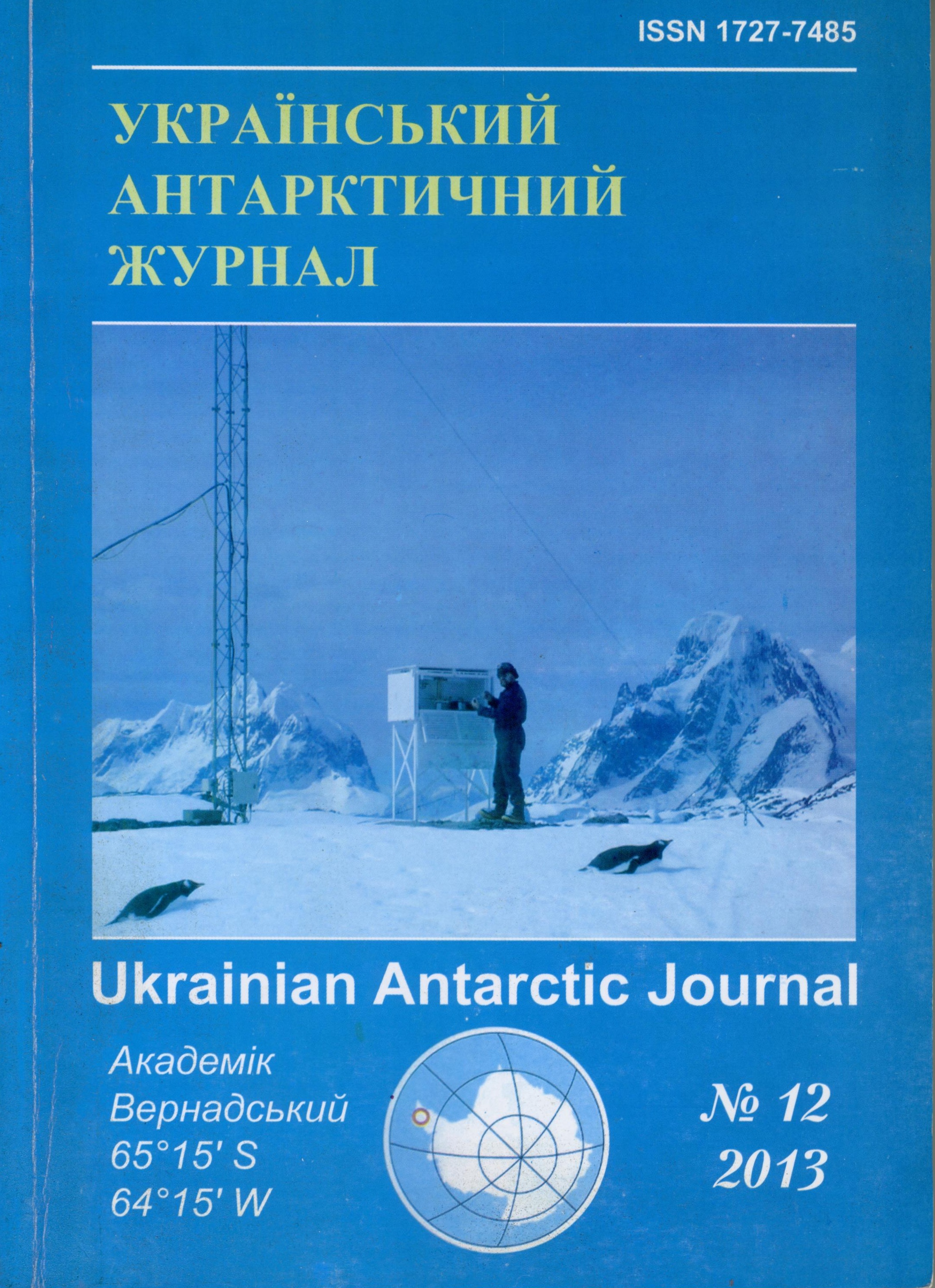
Відповідь фотосинтетичного апарату двох видів Deschampsia з різним ареалом розповсюдження на абіотичний стрес
- Deschampsia,
- UV-B radiation,
- carotenoids,
- glycolipids
Анотація
Deschampsia antarctica (ендемік Антарктичного регіону) і Deschampsia caespitosa (представник регіонів помірного клімату) – два види рослин родини Poaceae. Досліджували вплив УФ-В випромінювання та Н2О2 на фотосинтетичний апарат цих рослин. УФ-В випромінювання викликало деградацію хлорофілу а та β-каротину в листках рослин двох видів Deschampsia. Вміст галактоліпідів в листках обох видів в умовах УФ-В випромінювання суттєво варіював, але спостерігався відносно стабільний вміст сульфохіновозилдіацилгліцеролу. УФ-В випромінювання викликало неістотне зниження рівня окиснення пулу QA в листках D. аntarctica та підвищення цього показника в листках D. caespitosa. Хоча дія УФ-В викликала невелике зниження нефотохімічного гасіння в листках D. сaespitosa, квантова ефективність ФС ІІ залишалася незмінною. Співвідношення між мономерними та олігомерними формами LHC II (LHCP1/LHCP3) у фотосинтетичному апараті опромінених рослин обох видів Deschampsia підвищувалося особливо суттєво для D. сaespitosa. Обробка рослин H2O2 викликала несуттєве зниження активності СОД в обох видів. Пігментний склад характеризувався підвищенням вмісту каротиноїдів у листках рослин D. antarctica та вмісту хлорофілу а у обох видів. Вміст гліколіпідів у листках був стабільним, а вміст СХДГ дещо підвищувався після обробки H2O2 рослин D.antarctica.
Посилання
- Anderson, J.M. (1980). P-700 content and polypeptide profile of chlorophyll-protein complexes of spinach and barley thylakoids. Biochim. et Biophys. Acta, 591(1), 113–126.
- Arnon, D. (1949). Copper enzymes in isolated chloroplasts. Polyphenol oxidase in Beta Vulgaris. Plant Physiol., 24(1), 1-15.
- Barber, J. (1994). Molecular basis of the vulnerability of Photosystem II to damage by light. Aust J Plant Physiol., 22(2), 201–208.
- Barber, J., & Gounaris, K. (1986). What role does sulpholipid play within the thylakoid membrane? Photosynth Res., 9(1–2), 239–249.
- Bugos, R.C., Hieber, A.D., & Yamamoto, H.Y. (1998). Xanthophyll cycle enzymes are members of the lipocalin family, the first identified from plants. The Journal оf Biol.Chem., 273(25), 15321–15324.
- Bugos, R.C., & Yamamoto, H.Y. (1996). Molecular cloning of violaxanthin de-epoxidase from romaine lettuce and expression in Escherichia coli. Proceedings of the National Academy of Science of USA, 93(13), 6320–6325.
- De Vitry, C., Ouyang, Y., Finazzi, G., Wollman, F.-A., & Toivo, K. (2004). The chloroplast rieske iron-sulfur protein at the crossroad of electron transport and signal transduction.The Journal of Biol.Chem., 279(43), 44621–44627.
- Edge, R., McGarvey, D.J., & Truscott, T.G. (1997). The carotenoids as antioxidants. J. Photochem. Photobiol., 41(3), 189–200.
- Finazzi, G., Johnson, G.N., Osto, L.D., Joliot, P., Wollman, F.-A., & Bassi, R. (2004). A zeaxanthin-independent nonphotochemical quenching mechanism localized in the photosystem II core complex. PNAS, 101(33), 12375–12380.
- Foyer, C.H., Lelandais, M., & Kunert, K.J. (1994). Photooxidative stress in plants. Physiol. Plant, 92(4), 696–717.
- Gasser, A., Raddatz, S., Radunz, A., & Schmid, G.H. (1999). Comparative immunological and chemical analysis of lipids and carotenoids of the D1-peptide and of the light-harvesting-complex of photosystem II of Nicotiana tabacum. Z. Naturforsch, 54(3-4), 199–208.
- González-Rodríguez, A.M., Tausz, M., Wonisch, A., Jiménez, M.S., Grill, D., & Morales, D. (2001). The significance of xanthophylls and tocopherols in photo-oxidative stress and photoprotection of three Canarian laurel forest tree species on a high radiation day. Journal of Plant Physiol., 158(12), 1547–1554.
- Hager, A., & Holocher, K. (1994). Localisation of the xanthophyll-cycle enzyme violaxanthin de-epoxidase within the thylakoid lumen and abolition of this mobility by a (light-dependent) pH decrease. Planta, 192(4), 581–589.
- Havaux, M., & Kloppstech, K. (2001). The protective functions of carotenoid and flavonoid pigments against excess visible radiation at chilling temperature investigated in Arabidopsis npq and tt mutants. Planta, 213(6), 953–966.
- Hideg, É., Barta, C., Kálai, T., Vass, I., Hideg, K., & Asada, K. (2002). Detection of singlet oxygen and superoxide with fluorescent sensors in leaves under stress by photoinhibition or UV radiation. Plant and Cell Physiol., 43(10), 1154–1164.
- Hincha, D.K. (2003). Effects of calcium-induced aggregation on the physical stability of liposomes containing plant glycolipids. Biochim. Biophys. Acta, 1611(1–2), 180–186.
- Joyard, J., Mareshal, E., Miege, C. (1998). Structure, distribution and biosynthesis of glycerolipids from higher plant chloroplasts. In: Siegenthaler, P.A., Murata, N. (Eds.), Lipids in Photosynthesis: Structure, Function and Genetics. Advances in Photosynthesis, Kluwer Acad. Publ., Dordrecht.
- Kean, E.L. (1968.) Rapid sensitive spectrophotometric method for quantitative determination of sulfatides. Journal of Lipid Research, 9(3), 314–327.
- Kenrick, J., & Bishop, D. (1986). The fatty acid composition of phosphatidylglycerol and sulfoquinovosyl diacylglycerol of higher plants in relation to chilling sensitivity. Plant Physiol., 81(4), 946–948.
- Latowski, D., Åkerlund, H.-E., & Strzałka, K. (2004). Violaxanthinde-epoxidase, the xanthophyll cycle enzyme, requires lipid inverted hexagonal structures for its activity. Biochem., 43(15), 417–420.
- Latowski, D., Kostecka-Gugala, A., & Strzalka, K. (2003). Effect of the temperature on violaxanthin de-epoxidation: Comparison of the in vivo and model systems. Russian J of Plant Physiol., 50(2), 173–177.
- Lee, A.G. (2000). Membrane lipids: it’s only a phase. Current Biology, 10(10), 377–379.
- Lim, B.P., Nagao, A., Terao, J., Tanaka, K., Suzuki, T., & Takama, K. (1992). Antioxidant activity of xanthophylls on peroxyl radical-mediated phospholipid peroxidation. Biochim. Biophys. Acta, 1126(2), 178–184.
- Livn, A., & Packer, E. (1969). Partial resolution of the enzymes catalyzing photophosphorylation. V. Interaction of coupling factor I from chloroplasts with ribonucleic acid and lipids. J. of Biol. Chem., 244(5), 1332–1338.
- Madronich, S., McKenzie, R.L., Björn, L.O., & Caldwell, M.M. (1998). Changes in biologically active ultraviolet radiation reaching the Earth’s surface. J. Photochem. Photobiol., 46(1), 5–19.
- Menikh, A., & Fragata, M. (1993). Fourier transform infrared spectroscopic study of ion binding and intramolecular interactions in the polar head of digalactosyldiacylglycerol. Eur. Biophys. J., 22(4), 249–258.
- Merzlyak, M.N. (1978). Densimetric determination of carotenoids in plants in thin layers of “Silufol” plates. Nauchnye doclady Vyshey shkoly. Biologicheskie nauki, 1, 134–138.
- Middleton, E.M., & Teramura, A.H. (1993). The Role of Flavonol Glycosides and Carotenoids in Protecting Soybean from Ultraviolet-B Damage. Plant Physiol., 103(3), 741–752.
- Müller-Moulé, P., Havaux, M., & Niyogi, K.K. (2003). Zeaxanthin deficiency enhances the high light sensitivity of an ascorbate-deficient mutant of Arabidopsis. Plant Physiology, 133(2), 748–760.
- Murata, N., & Siegenthaler, P.A. (1998). Lipids in photosynthesis: an overview. In: P.A. Siegenthaler, N. Murata (Eds.), Lipids in Photosynthesis: Structure, Function and Genetics. Advances in Photosynthesis. KluwerAcad. Publ., Dordrecht.
- Musil, C.F., Chimphango, S.B.M., & Dakora, F.D. (2002). Effects of elevated ultraviolet-B radiation on native and cultivated plants of Southern Africa. Annals of Bot., 90(1), 127–137.
- Noctor, G., & Foyer, C.H. (1998). Ascorbate and glutathione: keeping active oxygen under control. Annu. Rev. Plant. Physiol. Plant. Mol. Biol., 49, 249–279.
- Palozza, P., & Krinsky, N.L.(1992). Antioxidant effects of carotenoids in vivoand in vitro: an overview. Methods in Enzymoiogy, 213, 403–420.
- Perez-Torres, E., García, A., Dinamarca, J., Alberdi, M., Gutiérrez, A., Gidekel, M., Ivanov, A.G., Huner, N.P.A., Corcuera, L.J., & Bravo, L.A. (2004). The role of photochemical quenching and antioxidants in photoprotection of Deschampsia antarctica. Functional Plant Biology, 31(7), 731–741.
- Pick, U., Gounaris, K., Weiss, M., & Barber, J. (1985). Tightly bound sulfolipids in chloroplast CF0-CF1. Biochim Biophys Acta, 808(3), 415–420.
- Rockholm, D.C., & Yamamoto, H.Y. (1996). Violaxanthin deepoxidase. Purification of a 43-Kilodalton Lumenal Protein from Lettuce by Lipid-Affinity Precipitation with Monogalactosyl-diacylglyceride.Plant Physiol., 110(2), 697–703.
- Ruban, A.V., Philip, D., Young, A.J., & Horton, P. (1997). Carotenoid dependent oligomerisation of the major chlorophyll a/blight-harvesting complex of Photosystem II of plants. Biochem., 36(6), 7855–7859.
- Sakaki, T. (1998). Responses of plant metabolism to air pollution and global change. In: L.J., de Kok, I. Stulen (Eds.), Backhuys Publishers. The Netherlands.
- Sakaki, T., Ohnishi, J., Kondo, N., & Yamada, M. (1985). Polar and neutral lipid changes in spinach leaves with ozone fumigation: triacylglycerol synthesis from polar lipids. Plant and Cell Physiol., 26(2), 253–262.
- Sakaki, T., Saitol, K., Kawaguchi, A., Kondo, N., & Yamada, M. (1990). Conversion of monogalactosyl-diacylglycerols to triacylglycerols in ozone-fumigated spinach leaves. Plant Physiol., 94(2), 766–772.
- Sakaki, T., Tanaka, K., & Yamada, M. (1994). General metabolic changes in leaf lipids in response to ozone. Plant and Cell Physiol., 35(1), 53–62.
- Siefermann, D., & Yamamoto, H.Y. (1975). Light-induced deepoxidation of violaxanthin in lettuce chloroplasts. IV. The effects of electron-transport conditions on violaxanthin availability. Biochim. Biophys. Acta, 387(1), 149–158.
- Sielewiesiuk, J., Matula, M., Gruszecki, W.I. (1997). Photo-oxidation of chlorophyll ain digalactosyldiacyl-glycerol liposomes containing xanthophyll pigments: indication of a special photoprotective ability of zeaxanthin. Cell Mol Biol Lett., 2(1), 59–68.
- Spotts, R.A., Lukezic, F.L., & Lacasse, L. (1975). The effect of benzimrdazole, cholesterol, and a steroid inhibitor on leaf sterols and ozone resistance of bean. Phytopathol., 65(1), 45–49.
- Steel, C.C., & Keller, M. (2000). Influence of UV-B radiation on the carotenoid content of Vitis vinifera tissues. Biochemical Society Transactions, 28(6), 883–885.
- Telfer, A., De Las Rivas, J., & Barber, J. (1991). β-carotene within the isolated photosystem II reaction centre: photooxidation and irreversible bleaching of this chromophore by oxidised P680. Biochim Biophys. Acta, 1060(1), 106–114.
- Telfer, A., Dhami, S., Bishop, S.M., Phillips, D., & Barber, J. (1994). β-Carotene quenches singlet oxygen formed by isolated photosystem II reaction centers. Biochem., 33(8), 14469–14474.
- Tomlinson, H., & Rich, S. (1973). Anti-senescent compounds reduce injury and steroid changes in ozonated leaves and their chloroplasts. Phytopathology, 63(7), 903–906.
- Trevathan, L.E., Moore, L.D., & Orcutt, D.M. (1979). Symptom expression and free sterol and fatty acid composition of flue-cured tobacco plants exposed to ozone. Phytopathol., 69(6), 582–585.
- UNEP. (1998). UNEP Environmental effects of ozone depletion: 1998 Assessment, 1-209.
- Van Kooten, O., & Snel, J.F.H. (1990). The Use of Chlorophyll Fluorescence Nomenclature in Plant Stress Physiology. Photosynth. Res., 25, 147–150.
- Webb, M.S., & Green, B.R. (1991). Biochemical and biophysical properties of thylakoid acyl lipids. Biochim. Biophys. Acta, 1060(2), 133–158.
- Whitaker, B.D., Lee, E.H., & Rowland, R.A. (1990). EDU and ozone protection: Foliar glycerolipids and steryl lipids in snapbean exposed to O3. Physiol. Plant., 80(2), 286–293.
- Xiong, F.S., & Day, T.A. (2001). Effect of solar ultraviolet-B radiation during springtime ozone depletion on photosynthesis and biomass production of Antarctic vascular plants. Plant Physiol., 125(2), 738–751.
- Yakovenko, G.M., & Mihno, A.I. (1971). Method of isolation and separation lipids and chloroplasts by types. Fiziol. i biochim. kult. rast., 3(6), 651–656.
- Yamamoto, H. (1980). High speed quantitative assey on TLC (HPTLC) plates. In: W. Bertch, R. Raser (Eds.), Instrumental HPTLC. New York.
- Yamamoto, H.Y., Chenchin, E.E., & Yamada, D.K. (1974). Effect of chloroplast lipids on violaxanthin de-epoxidase activity. In: Avron, M. (Ed.), Proceedings of the Third International Congress on Photosynthesis. Elsevier Scientific, Amsterdam, The Netherlands.
- Yamamoto, H.Y., & Higashi, R.M. (1978). Violaxanthin deepoxidase. Lipid composition and substrate specificity. Arch. Biochem. Biophys., 190(2), 514–522.
- Zill, L., & Harmon, E. (1962). Lipids of photosynthetic tissue. I. Salicilic acid chromatography of the lipids from whole leaves and chloroplasts. Biochem. Biophys. Acta, 57(1), 573–575.

